




881
Articles published




Join thousands of students achieving their dreams with expert-led courses.
Join free and unlock everything on EasyShiksha.
Takes less than 3 seconds — no password needed
Please enter a valid email address.
Checking your email…
Almost done — just verify your number so we can send course updates and certificates.
Used only for verification & updates
Incorrect OTP. Please try again.
Takes less than 10 seconds
Please select your country.
Please select your state.


Medical imaging plays an important role in modern healthcare by helping medical professionals examine internal structures of the human body and support the diagnosis and monitoring of different health conditions. Technologies such as X-ray, computed tomography (CT), magnetic resonance imaging (MRI) and ultrasound are used in different clinical situations to produce images that assist medical assessment.
The Bachelor of Radio Imaging Technology (BRIT) is an undergraduate programme category associated with medical imaging, radiological technology and diagnostic procedures. It introduces students to scientific concepts, imaging equipment, patient positioning, image quality and safety practices. The programme may suit learners who are interested in healthcare technology and practical work in diagnostic environments.
Students considering this field should understand that programme names, course structures, eligibility conditions, clinical training and professional requirements can vary between institutions. Exploring the BRIT programme category on EasyShiksha can help students begin reviewing available academic options.
Bachelor of Radio Imaging Technology, commonly abbreviated as BRIT, is an undergraduate-oriented area of healthcare education focused on medical imaging and radiological technology.
The field brings together knowledge of human anatomy, basic medical sciences, imaging principles, equipment operation, patient care and radiation safety. Students may learn how different imaging methods work, how patients are prepared for procedures and how suitable images are obtained under established clinical protocols.
Medical imaging is not limited to operating equipment. It also requires an understanding of patient communication, accurate identification, positioning, safety procedures, image quality and professional responsibilities.
The exact qualification awarded and the scope of training depend on the individual programme and institution. Students should therefore examine the programme curriculum rather than assuming that every course listed under BRIT offers identical training.
Medical imaging supports several areas of healthcare. Doctors may use diagnostic images to investigate injuries, examine organs, monitor certain diseases or assess changes in a patient's condition.
The work involves coordination between radiologists, medical radiation technologists, medical physicists, nurses and other healthcare professionals. Each member of the team has responsibilities based on their education, training and professional role.
Studying BRIT may help students understand this multidisciplinary environment. It can introduce them to the relationship between medical science, technology and patient care.
Students interested in exploring other healthcare qualifications can also browse the EasyShiksha University Programmes Directory to review different programme categories.
The curriculum of a BRIT programme may include theoretical subjects, laboratory learning, practical demonstrations and supervised clinical exposure. The precise subject list differs between institutions.
Anatomy introduces students to the structure of the human body, including bones, muscles, organs and body systems. Physiology explains how these systems function.
This knowledge helps learners understand anatomical positioning and the structures visible in medical images. It also provides a foundation for understanding how imaging procedures relate to different clinical requirements.
Radiographic physics introduces the scientific principles behind X-ray production and image formation. Students may learn about radiation, equipment components, exposure factors and the relationship between technical settings and image quality.
A basic understanding of physics is useful when studying how imaging equipment produces diagnostic images.
Radiography is associated with producing images of internal body structures using X-rays. Students may learn about imaging procedures, patient positioning, equipment components and image evaluation.
Practical training may introduce learners to the steps involved in preparing patients and following the required procedure. Actual responsibilities during clinical work depend on the student's training and supervision.
Computed tomography, commonly called CT, uses X-rays and computer processing to create cross-sectional images of the body.
A programme may introduce students to CT equipment, scanning principles, image reconstruction and patient preparation. The depth of CT-related learning depends on the curriculum and available clinical facilities.
Magnetic resonance imaging, or MRI, uses magnetic fields and radiofrequency signals to produce images of internal body structures.
Students may study the basic principles of MRI, patient screening, safety considerations and imaging procedures. MRI has distinct safety requirements, so learners must understand the importance of following institutional protocols.
Ultrasound uses high-frequency sound waves to create images of internal structures. It is used in several clinical areas and does not rely on ionising radiation.
Students may receive an introduction to ultrasound principles, equipment components and the role of ultrasound in diagnostic imaging, depending on the programme structure.
Radiation safety is an essential area of medical imaging education. Students may learn about exposure control, protective equipment, safe working practices, patient identification and institutional safety procedures.
International guidance highlights the importance of appropriate education and training for healthcare professionals involved in medical uses of radiation. Students should therefore pay close attention to the radiation protection component of any programme they consider.
Imaging procedures often involve direct interaction with patients. Learners may study patient preparation, communication, positioning assistance, infection-control practices and professional conduct.
Clear communication can help patients understand the procedure and follow the instructions provided by authorised healthcare professionals.
Practical learning is an important consideration when evaluating a medical imaging programme. Classroom knowledge alone may not provide sufficient preparation for understanding the working environment of a diagnostic imaging department.
Depending on the institution, practical components may include:
Demonstrations of radiographic equipment and imaging procedures.
Learning about patient positioning and preparation.
Understanding image quality and basic image evaluation.
Laboratory activities related to imaging principles.
Observation of diagnostic imaging workflows.
Supervised clinical exposure, where included in the programme.
Students should ask institutions about the availability of imaging laboratories, clinical affiliations, practical assessments, supervision and internship arrangements.
It is important to distinguish between observing a procedure, practising under supervision and independently performing a professional responsibility. Students should not assume that admission to a programme automatically authorises them to perform every imaging procedure.
Eligibility requirements depend on the institution, the programme structure and the admission rules applicable to the academic year.
Common requirements may include:
Completion of Class 12 or an equivalent qualification.
A science background, where specified by the institution.
Physics, Chemistry and Biology or another prescribed subject combination.
Meeting the minimum marks required by the institution.
Satisfying any applicable entrance examination or selection criteria.
Submitting the required academic and identity documents.
Some institutions may have additional requirements related to age, medical fitness or admission procedures.
Students should verify the current eligibility criteria directly from the individual programme information before submitting an application. The BRIT programme listing on EasyShiksha provides a starting point for exploring the category.
The duration and academic structure of BRIT programmes may vary by institution. Some programme listings describe a three-year structure, but students should verify the duration of the specific course they intend to pursue.
The study structure may include:
Semester-based academic learning.
Classroom lectures and assignments.
Laboratory or equipment-based practical sessions.
Internal and external assessments.
Clinical observation or supervised training, where prescribed.
Internship or other practical components, if required.
Before admission, students should confirm the total programme duration, semester structure, attendance requirements, examination pattern and any compulsory clinical training.
The admission process may involve merit-based selection, entrance examinations, counselling or another method prescribed by the institution.
Begin by identifying institutions that offer a Bachelor of Radio Imaging Technology or a related medical imaging qualification. Review the BRIT programme options on EasyShiksha to start your search.
Read the admission requirements carefully. Confirm the required Class 12 subjects, minimum marks and any additional conditions.
Examine the subjects, practical components, imaging technologies covered and clinical exposure offered by the institution.
Check whether admission is based on academic merit, an entrance examination, counselling, an interview or another selection process.
Fill out the application form with accurate personal, academic and contact information. Follow the institution's instructions for submission.
Depending on the institution, documents may include academic mark sheets, certificates, identity proof, photographs and other applicable records.
Before accepting admission, review the programme duration, qualification awarded, learning mode, practical training arrangements and applicable institutional requirements.
BRIT-related education may help learners develop a combination of technical, analytical and interpersonal skills.
Technical understanding: Students may develop familiarity with imaging equipment, radiographic principles and diagnostic procedures.
Anatomical knowledge: Understanding body structures can help learners interpret positioning requirements and imaging concepts.
Attention to detail: Medical imaging requires careful attention to patient identification, positioning, procedure instructions and image quality.
Patient communication: Students may develop the ability to communicate clearly and professionally with patients and healthcare teams.
Safety awareness: Learning about radiation protection, equipment safety and clinical protocols can support responsible professional practice.
Analytical thinking: Understanding imaging principles and image quality can encourage observation, problem-solving and technical reasoning.
These skills depend on the curriculum, practical experience, supervision and individual learning progress.
Career possibilities depend on the qualification awarded, the programme's practical components, employer requirements and applicable professional regulations.
Depending on their training and eligibility, graduates may explore opportunities in areas such as:
Diagnostic imaging departments.
Hospitals and healthcare facilities.
Medical imaging centres.
Radiography-related services.
Imaging equipment and technical support environments.
Healthcare education or research support, where suitably qualified.
The specific job title and responsibilities should always be checked against the qualification and applicable requirements. Completing a programme does not automatically confer eligibility for every regulated healthcare role.
Students should also understand the distinction between a radiologist, who is a medical doctor with specialist training, and a medical radiation technologist or radiographer, whose role is based on a different education and professional pathway.
Students who complete an eligible undergraduate qualification may consider further education in related areas. Possible academic directions can include postgraduate study in medical imaging, radiological technology, healthcare sciences, biomedical sciences or other relevant disciplines.
The availability of these options depends on the qualification, institution and entry requirements of the postgraduate programme.
Before selecting a higher-study route, students should verify whether their undergraduate qualification meets the required academic criteria. They should also review the subjects, practical experience and any additional conditions required for admission.
Choosing a medical imaging programme requires more than checking its name or advertised duration. Students should examine the academic and practical details before making a decision.
Consider the following points:
Curriculum coverage: Check whether the programme includes relevant subjects such as anatomy, radiographic physics, imaging principles, patient care and radiation protection.
Practical facilities: Understand what equipment, laboratories and practical learning opportunities are available.
Clinical exposure: Ask whether supervised clinical training is included and how it is organised.
Qualification details: Confirm the exact title of the qualification and the institution awarding it.
Eligibility and admission: Review the current admission requirements and selection method.
Professional requirements: Check the rules applicable to the professional roles you may wish to pursue after graduation.
Academic progression: Consider whether the qualification supports your intended higher-study plans.
The individual programme page for BRIT in Radio Imaging Technology can be used to review one listed programme. Students should independently verify current institutional and professional requirements before applying.
The Bachelor of Radio Imaging Technology is an undergraduate-oriented healthcare education pathway associated with medical imaging, radiographic techniques, diagnostic technology and patient care. It may introduce students to areas such as anatomy, radiographic physics, X-ray imaging, CT, MRI, ultrasound and radiation protection.
For students interested in combining healthcare knowledge with technology and practical learning, BRIT may be an academic option worth exploring. However, programme structures, eligibility, clinical exposure, qualification details and career requirements can differ between institutions.
Before applying, students should carefully review the curriculum, practical facilities, clinical training arrangements and professional requirements associated with their intended qualification. A well-informed decision can help learners choose a programme that aligns with their academic interests and long-term goals.
BRIT commonly refers to Bachelor of Radio Imaging Technology. It is an undergraduate-oriented healthcare education category associated with medical imaging, radiography and diagnostic technology.
Students may study anatomy, physiology, radiographic physics, X-ray imaging, CT, MRI, ultrasound, radiation protection, patient care and medical imaging procedures, depending on the institution.
Applicants commonly need Class 12 or an equivalent qualification. Required subjects, minimum marks and entrance examination conditions depend on the institution and its current admission rules.
The duration varies by institution and programme structure. Students should check the official course details to confirm the academic duration and any additional clinical or internship requirements.
Practical learning may include laboratory sessions, equipment demonstrations and supervised clinical exposure. Students should confirm the actual training facilities and arrangements offered by their chosen institution.
Depending on the curriculum, students may be introduced to X-ray, computed tomography, magnetic resonance imaging and ultrasound, along with related imaging principles and safety practices.
Depending on their qualification and applicable requirements, graduates may explore opportunities in diagnostic imaging departments, hospitals, medical imaging centres and related healthcare environments.
No. A radiologist is a medical doctor with specialist training. BRIT is associated with medical imaging technology education and a different professional pathway.
Eligible graduates may explore postgraduate education in medical imaging, radiological technology or related healthcare fields. Admission depends on the qualification and the requirements of the institution.
Students should review eligibility, curriculum, qualification details, practical facilities, clinical training, institutional requirements and the professional rules relevant to their intended career.
More News Click here










Discover thousands of colleges and courses, enhance skills with online courses and internships, explore career alternatives, and stay updated with the latest educational news..

Gain high-quality, filtered student leads, prominent homepage ads, top search ranking, and a separate website. Let us actively enhance your brand awareness.
Download EasyShiksha Mobile Apps from Android Play Store, Apple App Store, Amazon App Store, and Jio STB.
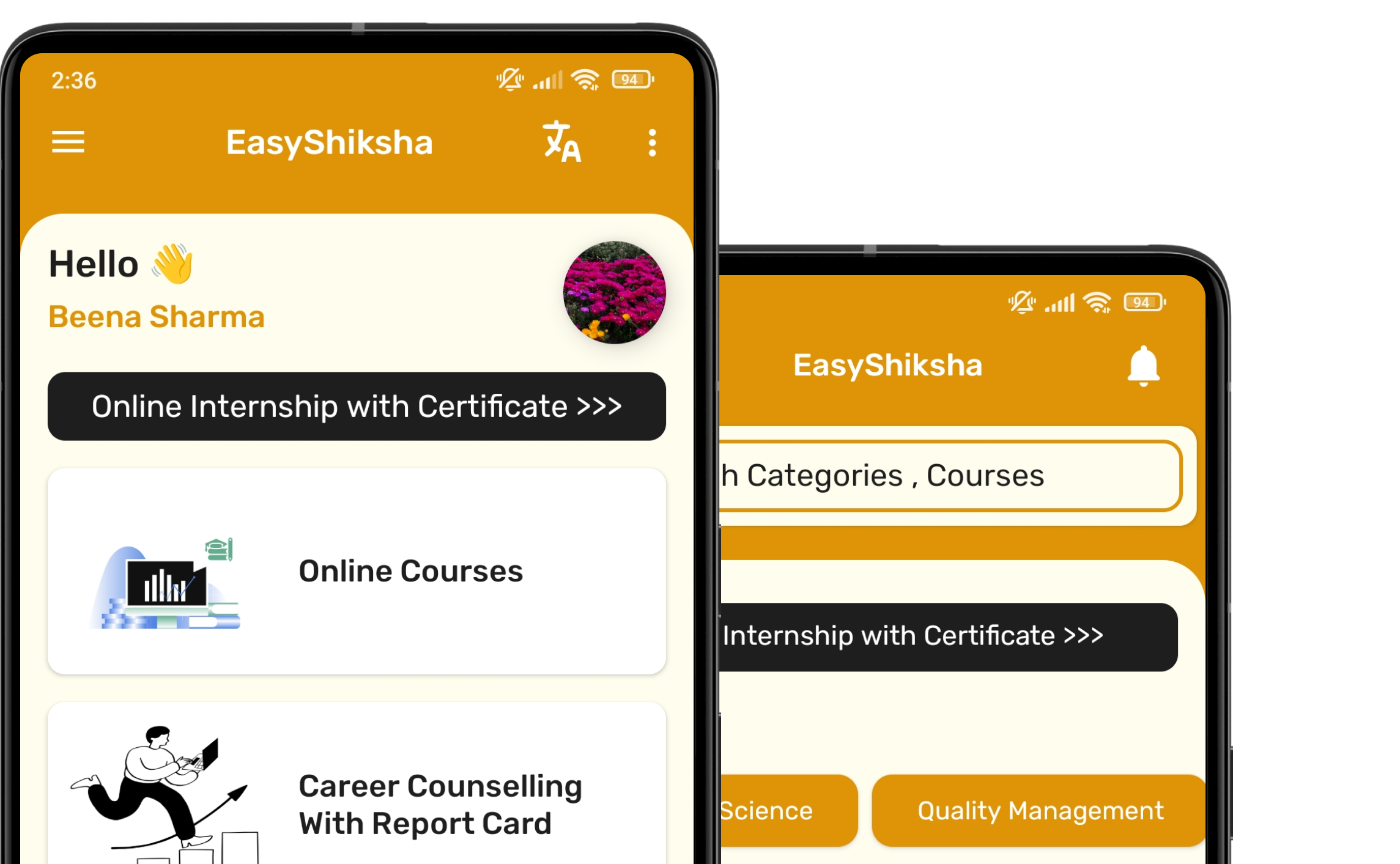


1 million+ learned on the EasyShiksha App
Trusted by Standards & Global Compliance Frameworks




Built under internationally recognized standards for quality, information security, educational excellence and business continuity.
ISO 9001:2015 | ISO/IEC 27001:2022 | ISO 21001:2018 | ISO 22301:2019 | DPIIT Recognized Startup | MSME Registered